Mastering Ultrasound: The Secret Lies in Your Grip
When performing an ultrasound, most veterinary professionals focus on image quality, machine settings, and anatomical accuracy. But what if a crucial...
1 min read
.jpg) Fred Gromalak, DVM
:
Jan 24, 2025 5:33:09 PM
Fred Gromalak, DVM
:
Jan 24, 2025 5:33:09 PM

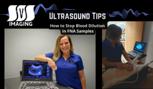
Fine needle aspirates (FNAs) are a cornerstone diagnostic tool in veterinary medicine, but one common frustration is blood contamination in the sample. This can dilute results and hinder cytological analysis.
Dr. Fred Gromalak from SVS Imaging shares a quick and effective tip to overcome this issue:
1️⃣ Attach the needle to your syringe and locate the area of interest.
2️⃣ Aspirate the lesion as usual. If blood fills the hub, stop.
3️⃣ Remove the needle and replace it with a new one.
4️⃣ Place your finger over the hub to create negative pressure.
5️⃣ Reattempt aspiration using the same technique.
This small adjustment significantly reduces blood contamination, allowing you to collect high-quality samples for more accurate diagnoses.
Master this simple trick and elevate your veterinary cytology game!
👉 Watch the video below:
Need Support? The SVS Imaging team offers mobile ultrasound services, ultrasound bootcamps, and virtual telemedicine consultations.
For more insights and training opportunities, visit our website or contact us today!
#VeterinaryUltrasound #UrethralImaging #VetMed #SVSImaging #DiagnosticUltrasound

When performing an ultrasound, most veterinary professionals focus on image quality, machine settings, and anatomical accuracy. But what if a crucial...

Struggling to ultrasound large dogs? Learn four essential tips from Dr. Fred Gromalak of SVS Imaging to improve your technique, from sedation to...

Accurately identifying the left adrenal gland in canine ultrasound imaging can sometimes be tricky due to the presence of nearby structures like...