Leveraging Multiple Specialties on Complex Cases

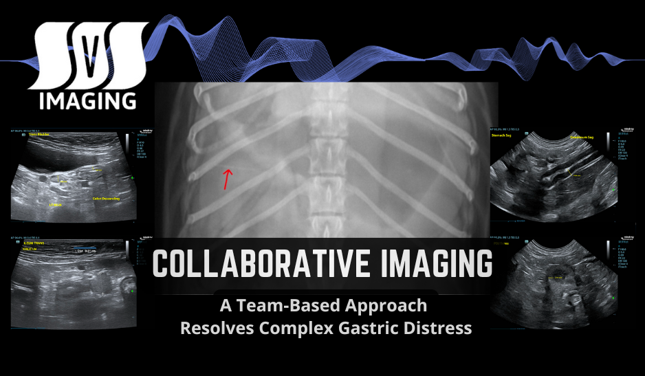
When facing a challenging gastrointestinal case, the power of collaboration cannot be overstated. In this recent case, a 1-year and 7-month-old spayed female Mini Dachshund presented at an emergency clinic with significant gastric distention, regurgitation, and concerns about a possible gastrointestinal obstruction.

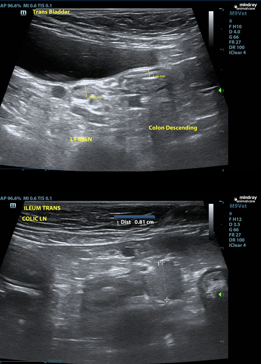
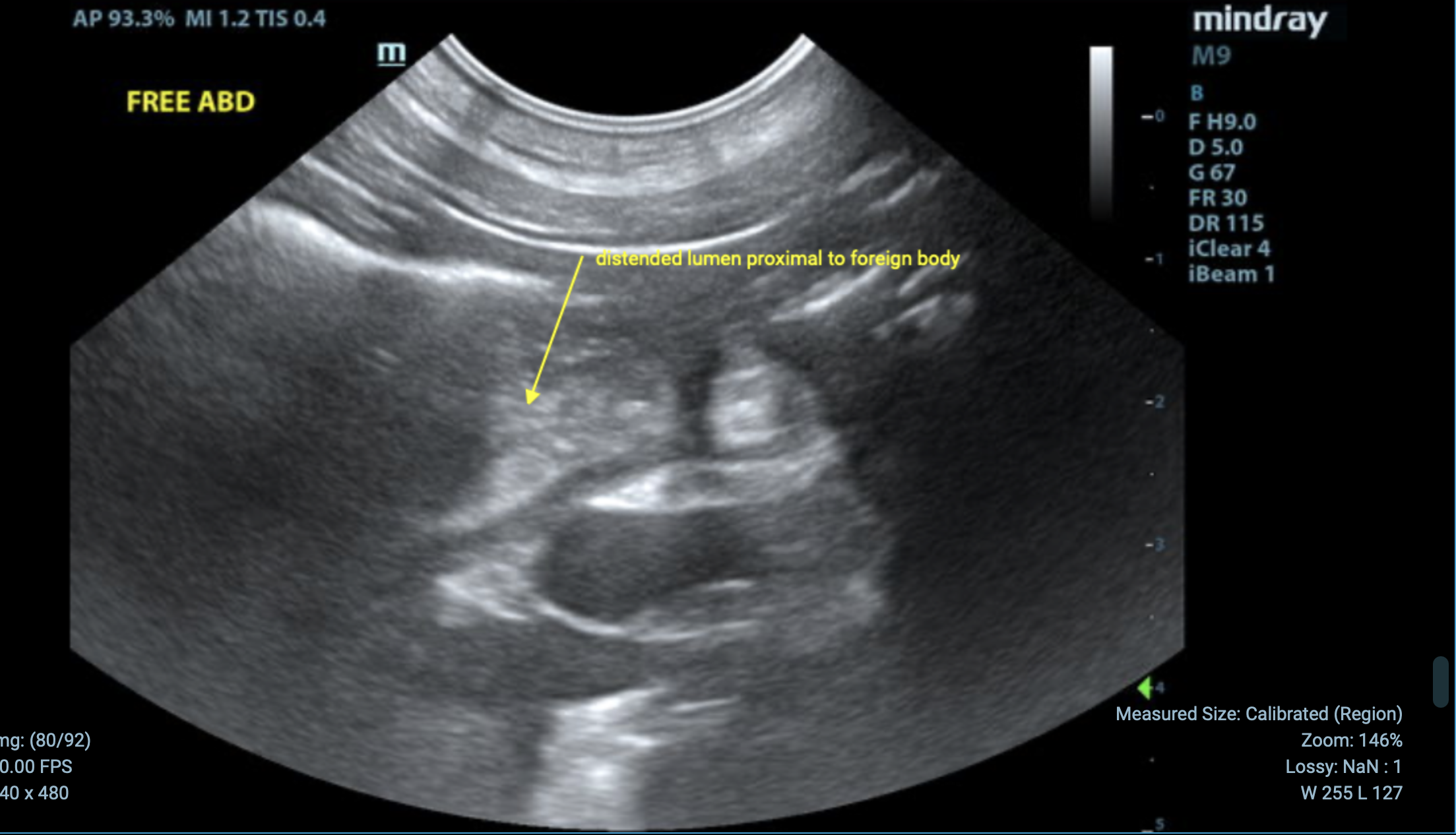
The patient had undergone an initial ultrasound exam the previous day, which revealed an ileal foreign body prompting a STAT multi-modality (ultrasound and radiograph) review by the SVS Imaging team. By leveraging the expertise of both a SVS Imaging radiologist and internist, we were able to provide a detailed and actionable care plan.

 
Radiologist's Observations (RADIOGRAPH):
RAD - Collaborative Imaging A Team-Based Approach Resolves Complex Gastric Distress

  • Gastric distention; rule out functional ileus/atony secondary to pain or inflammation (gastritis, ulceration, peritonitis, pancreatitis).

  • No gastric foreign material or outflow obstruction is identified on this study.

  • Nasogastric tube placement to reduce distention and discomfort is recommended. 

  • Continue medical management for nonspecific gastroenteritis.

  • Consider snap cPLI testing to assess for pancreatitis, even if sonographically normal.

  • If the patient continues to decline, consider barium study or abdominal CT.

INTERNIST's Perspective (ULTRASOUND):
 
STOMACH
  • The stomach contains a marked amount of hypoechoic fluid and gas, indicative of gastritis and gastric ileus. No obvious shadowing foreign body is identified.
  • The wall of the body of the stomach measures 0.4 cm in width, which is within normal limits.
  • The pylorus is visualized, there is no evidence of pyloric outflow obstruction at this time.
Stomach and Pylorus

Duodenum_Pancreas


DUODENUM
  • The duodenum is empty and normal in size, measuring 4.8 mm in width, with normal layering observed.
PANCREAS
  • The pancreas is isoechoic to the surrounding mesentery, with no abnormalities noted.

Ileum_Colon


  • Ileum
    • The ileum appears empty at this time, with the wall measuring 2.1 mm in width.
    • There is no evidence of the previously seen foreign material; it appears to have passed into the colon. However, this material is not visualized in the colon due to the presence of gas and feces.


  • Colon
    • The colon is empty at this time, with a normal wall thickness of 1.7 mm.


US - Trans Bladder - Ileum - Collaborative Imaging A Team-Based Approach Resolves Complex Gastric Distress


Urinary Bladder
  • The urinary bladder contains a small amount of anechoic urine with no bladder stones or masses detected. The bladder wall is diffusely normal in thickness.

LYMPH NODES
  • Several mildly enlarged medial iliac lymph nodes are observed on the left side, likely reactive or age-related. One representative lymph node measures 2.8 mm in width.
  • A colonic lymph node is also mildly enlarged, measuring 0.8 cm in width, appearing reactive and less likely due to neoplasia.

INTERNIST'S CONCLUSIONS
  • The previously identified ileal foreign body appears to have passed into the colon, with no current evidence of obstruction.
  • The enlarged lymph nodes are likely reactive due to the previous foreign body obstruction and do not appear neoplastic.
  • The stomach is diffusely enlarged with hypoechoic fluid, but no gastric foreign body or pyloric outflow obstruction is present at this time.
Prognosis
  • The prognosis appears good. With supportive care, the patient is expected to improve. However, if clinical signs persist, further diagnostic interventions such as endoscopy or surgery may be warranted.

Recommendations

Supportive Care
  • Continue supportive care, including monitoring of the patient’s stool for the passage of previously seen foreign material.
  • Consider placing a nasogastric tube to decompress the stomach and start feeding a liquid diet to facilitate gastric motility.
Diagnostic Testing
  • Submit a quantitative cPLI test to screen for possible occult pancreatitis, which may be contributing to the patient's gastritis.
Medications
  • Recommend prokinetics such as erythromycin at a dosage of 0.5 to 1 mg/kg, administered either orally or IV every 8 hours.
  • Initiate trickle feeding with a low-fat liquid diet to promote gastric motility.
Further Evaluation
  • If the patient does not show improvement with supportive care, consider endoscopy or surgical intervention for biopsies of the stomach to evaluate for inflammatory diseases such as inflammatory bowel disease.
Collaborative Approach in action

This case exemplifies the synergy between veterinary imaging and clinical

expertise. 

  • Multi-Modality Approach: Integrating ultrasound and radiographic findings.
  • Radiologist’s Precision: High-quality imaging and detailed analysis of subtle abnormalities.
  • Internist’s Insight: Synthesis of imaging findings with the patient’s clinical presentation to highlight differential diagnoses, including possible functional gastric outflow obstruction. Recommended supportive care to alleviate gastric stasis, including nasogastric tube placement, prokinetics, and ongoing diagnostics to rule out pancreatitis or delayed gastric emptying.
  • Outcome: A comprehensive care plan that empowered the referring veterinarian to confidently proceed with a clear treatment strategy. Early indications showed improvement, thanks to the combined efforts of SVS Imaging’s specialists and the referring veterinary emergency team.
    Sample Report - A Team-Based Approach Resolves Complex Gastric Distress


Want to learn more?

About SVS Imaging

Veterinary Teleradiology and Internal Medicine Consultations: Veterinarians worldwide can rely on our specialists' expertise in interpreting RADS, CT scans, MRIs, and ultrasound images, and providing you with accurate and comprehensive diagnoses. Click here to learn more.USA Teleradiology_ Accurate Interpretations by Certified Radiologists

USA Telemedicine_  Virtual Consult with a Specialist