.jpg) Fred Gromalak, DVM
Fred Gromalak, DVM
1 min read
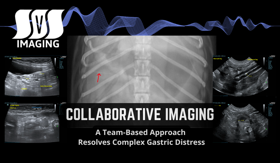
Collaborative Imaging: A Team-Based Approach Resolves Complex Gastric Distress
Leveraging Multiple Specialties on Complex Cases
As veterinarians, one of the most critical aspects of diagnosing abdominal issues is the ability to effectively interpret ultrasound images. Whether it’s dealing with gastrointestinal blockages or detecting hidden foreign bodies, ultrasound can offer invaluable insights into a pet’s condition.
In the video below, Dr. Fred Gromalak, DVM is going to through an emergency “foreign body hunt” using ultrasound images obtained by SVS Imaging Mobile Sonographer Kim Liedberg. This live demonstration shows how we identify and assess possible obstructions.
Now, let’s walk through the key steps to investigate a suspected foreign body in the gastrointestinal tract.

Our first focus is the urinary bladder, which is a helpful reference point for assessing abdominal structures.
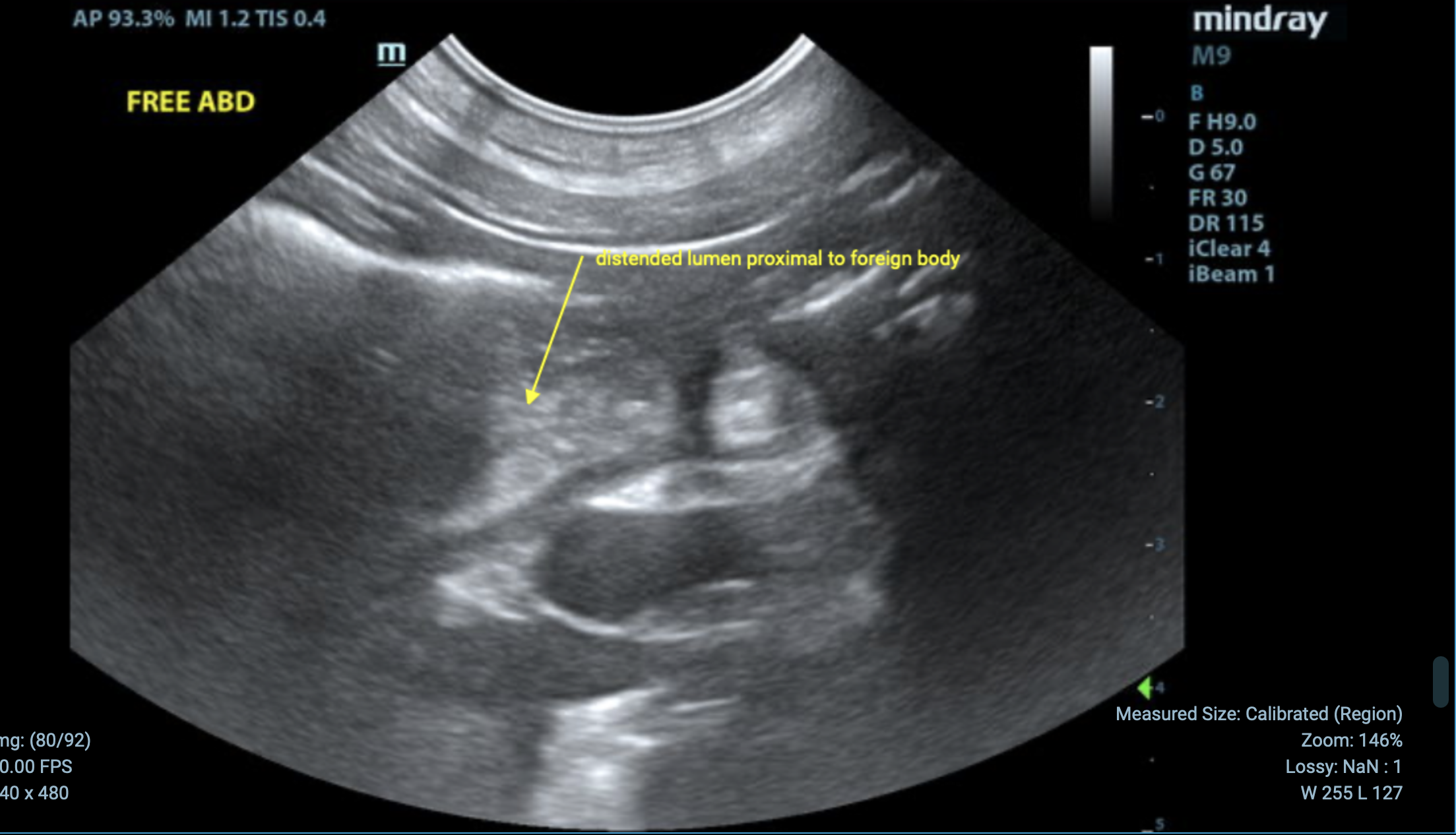
Next, we turn our attention to the small intestine. In this particular case, we notice small intestinal luminal dilation, which can be a sign of a potential obstruction.
Adjacent to the bladder, we see the descending colon. It’s crucial to observe whether there’s any foreign body in the gastrointestinal tract, and in this instance, no obvious obstruction is detected in the colon.
STOMACH
As we continue our exam, we observe the stomach, which shows mild distention, gas, and some loose ingesta (digested material). This suggests that the gastrointestinal system is attempting to process food but may be encountering some obstacles.

PYLOROUS
Evaluation of the pyloric antrum revealed no evidence of foreign material. Therefore there is no evidence of an upper gastrointestinal obstruction (i.e. pyloric outflow obstruction).


After reviewing all the structures and imaging, we notice a segment of the small intestine that stands out due to its dilation and suspicious appearance. While we don’t see clear evidence of a foreign body in this segment, the combination of luminal dilation, regional inflammation, and wall thickening raises strong suspicion for a foreign object blocking the intestine.

At this point in the exam, we’ve gathered enough evidence to recommend further exploration. While ultrasound can provide valuable clues, it’s essential to take a closer look and confirm the presence of a foreign body through more invasive procedures, such as surgery or endoscopy, if necessary.
Foreign bodies are a common issue in veterinary medicine, and detecting them early is key to preventing complications. By using ultrasound to closely examine the abdomen, we can pinpoint areas of concern and take action quickly, ensuring the best outcome for our patients. By following the steps outlined in this blog post, you can see how we systematically assess the abdominal organs and use ultrasound to detect abnormalities that might otherwise go unnoticed.
The “foreign body hunt” isn’t always straightforward, but with a careful and thorough approach, we can find and address issues early on, helping our patients feel better faster.
Stay tuned for more insights on veterinary diagnostics, and remember: when it comes to foreign bodies, always trust your instincts and keep hunting for the clues that will lead you to the right diagnosis!

1 min read
Leveraging Multiple Specialties on Complex Cases
1 min read
When you first step onto a golf course, the swing feels unnatural. You’re told to hold the club a certain way, position your feet just right, and...
1 min read
Struggling to ultrasound large dogs? Learn four essential tips from Dr. Fred Gromalak of SVS Imaging to improve your technique, from sedation to...