When performing bladder ultrasounds, it's easy to focus primarily on sagittal and transverse views. However, the urethra is a critical area where pathology often hides. In this post, Dr. Fred Gromalak from SVS Imaging discusses a real case study, demonstrates essential imaging techniques, and highlights why thorough urethral imaging is essential for accurate diagnosis.
Case Study: Chronic Hematuria in a 14-Year-Old Dachshund
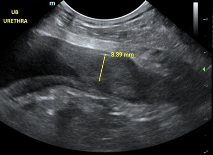
A 14-year-old female spayed dachshund presented with chronic hematuria and a lack of response to antibiotics. During the ultrasound examination, asymmetrical thickening was observed at the urethra-bladder junction, measuring up to 0.83 cm.
🔑 Key Takeaways:
-
Possible causes include transitional cell carcinoma or nonspecific inflammation/urethritis.
-
A screening BRAF assay was recommended for further evaluation.
-
If non-diagnostic, fine-needle aspiration would be the next step to assess cellular composition and confirm or rule out neoplasia.
Download the full case report
The Technique: Getting the Right Urethral View
In a follow-up demonstration, Dr. Gromalak explains how to optimize your ultrasound technique to visualize the urethra effectively:
-
Imagine showing a watch on your wrist to the patient's head.
-
Perform a twist and tilt maneuver to align your probe correctly.
-
Look for a triangular shape on the ultrasound screen, which indicates proper urethral alignment.
-
A round shape suggests you're moving into the bladder, while a pointed triangle ensures you're tracking the urethra.
Why This Matters:
Proper imaging of the urethra can mean the difference between an early diagnosis and a missed pathology. Whether it's neoplasia, inflammation, or structural abnormalities, precision matters.
Watch the Full Demonstration:
Don't overlook the urethra during bladder ultrasounds. Adopting these techniques can significantly enhance your diagnostic accuracy and improve patient outcomes.
Need Support? The SVS Imaging team offers mobile ultrasound services, ultrasound bootcamps, and virtual telemedicine consultations.
For more insights and training opportunities, visit our website or contact us today!
#VeterinaryUltrasound #UrethralImaging #VetMed #SVSImaging #DiagnosticUltrasound
.jpg) Fred Gromalak, DVM
Fred Gromalak, DVM