Veterinary Cardiology Echocardiography Training Course
Canine Cardiac Imaging in an Intensive 3-Day Hands-On Course.
Next Session: Nov 30 - Dec 2, 2026
Limited to 9 participants per session
Small Class Size
Limited to 9 participants for personalized attention.
Hands-on Experience
with guidance from expert cardiologists and sonographers.
Enhanced Confidence
Perform diagnostic-quality echocardiograms with confidence
3-day intensive, hands-on
Waukesha, WI
20.5 CE Credits
Build Skills That Translate to Better Images
This intensive three-day course is designed to help veterinarians and veterinary technicians:
Perform complete canine echocardiograms.
Consistently obtain diagnostic-quality imaging.
Submit studies suitable for board-certified cardiologist interpretation.
Hands-on training with real canine patients under the guidance of a veterinary cardiologist and experienced sonographersensures you gain confidence and proficiency.
Is This Course Right for You?
This course is ideal for veterinarians and technicians with:
12–24 months of clinical experience in small animal practice.
Prior exposure to diagnostic imaging, including abdominal or thoracic ultrasound.
Familiarity with common canine cardiac diseases such as degenerative mitral valve disease, dilated cardiomyopathy, PDA, VSD, or PS.
Pre-Course Requirements:
Access to a cardiac-capable ultrasound machine, ECG, and echocardiography table.
Basic knowledge of cardiac anatomy and function.
Completion of pre-course assessment including reading materials and image submission.
What you'll Learn:
Understand indications, limitations, and principles of canine echocardiography.
Properly position canine patients for transthoracic echocardiography.
Identify and obtain all standard echocardiographic views, including: - Right parasternal long-axis (4-chamber, 5-chamber) - Right parasternal short-axis (mitral, papillary muscle, chordae, aortic valve, pulmonary artery) - Left apical views (4-chamber, 5-chamber) - Subcostal and cranial abdominal views (as indicated)
Optimize image acquisition for 2D, M-mode, and Doppler (PW, CW, color) studies.
Document studies to meet diagnostic standards for cardiologists.
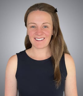
Dr. Morey, originally from Nairobi, Kenya, earned her veterinary degree at the University of Edinburgh. After completing a rotating internship in Denver and a cardiology specialty internship in Seattle, she completed her cardiology residency in Columbia, Missouri, achieving board certification in 2023. She then served as a lecturer and co-head of the cardiology service at the Royal Veterinary College from 2023 through December 2025.
Register Now and Build Skills That Stay With You
Ready to build confidence, sharpen your skills, and bring high-quality ultrasound diagnostics back to your hospital?
Select your desired training date below to sign up. Space is limited!
• Foundations & echocardiographic anatomy • Live demonstrations: patient positioning & probe handling • Hands-on practice: right parasternal long & short-axis views
Day 2
Left Parasternal Imaging
• Common pitfalls & corrections • Hands-on practice: left apical 4 & 5-chamber, cranial views • Introduction to Doppler imaging
Day 3
Integrating Color and Velocity
• Doppler review and clinical applications • Hands-on practice: M-mode, color Doppler, PW and CW assessments • Complete your first full diagnostic echocardiogram
Four years ago, I learned to scan major abdominal organs as a beginner. Returning for a refresher course, I focused on the gastrointestinal tract and smaller structures. Familiarity with the protocol helped me learn advanced skills quickly, and I gained valuable new knowledge. The staff is supportive and passionate about teaching, and I’m excited to apply these ultrasound skills in general practice.
Sierra
Ultrasound Lab Attendee
Best CE I've Ever had! I'm scanning with confidence, thanks to Fred and his team. Excited to share this course with SVP and return for Echo training!
Mark
Ultrasound Lab Attendee
This was a very helpful course and improved my skills at some of the finer points of ultrasound. The repetitive work was crucial at solidifying skills.
Jeff
Lab Attendee
Class size was nice, instructors very helpful with guidance
Ruth
Lab Attendee
I had a great experience and felt I took away valuable and useful technique/protocol from this camp.
Chloe
Ultrasound Attendee
The three day course was a really awesome training experience. Lots of hands on training packed into a fun, relaxed, and efficient 3 days. Highly recommend.
Adam
Lab Attendee
I came to SVS imaging to further my ultrasound skills and be able to perform diagnostics ultrasounds. I definitely learned more and got more valuable skills to take back to my clinic. Everyone at the COVE was so willing to teach and share their knowledge.
Michayla
Lab Attendee
Such an amazing and informative course with outstanding staff present. Tons of knowledge learned and will take back to my clinic.