.jpg) Fred Gromalak, DVM
Fred Gromalak, DVM
1 min read
Exploring the World of Veterinary Mobile Ultrasound
Road warriors of the Veterinary Field In the dynamic realm of veterinary medicine, there's a hidden gem that's gaining significant traction - the...
In recent years, the demand for in-house veterinary diagnostics has led to general veterinary practices investing in advanced equipment like CT scanners. CT offers a wealth of information for diagnosing various conditions, but a new player in the CT market, known as Cone Beam CT (CBCT) or flat panel CT, is gaining prominence due to its compact size, lower radiation emissions, reduced power requirements, and high-quality imaging of hard bony structures.


However, while this innovative technology is on the rise, it comes with some crucial considerations. Let's explore the key differences between traditional CT and CBCT.
Traditional CT vs. CBCT
Power: Traditional CT relies on high-output electricity, requiring a 3-phase power supply. In contrast, CBCT typically operates at peak voltages of 60-120 kV, drawing power similar to that of a standard x-ray unit.
Beam Shape: Traditional CT employs a fan-shaped x-ray beam, capturing data in a thin layer and producing a single-slice image per scan. CBCT, on the other hand, emits a cone-shaped x-ray beam, capturing multiple projections from different angles to create the image.
Shielding Requirements and Radiation Exposure: CBCT, with its lower power and horizontal radiation emission, demands less shielding for the room compared to traditional CT.
Originally developed for dental professionals due to its advantages, such as mobility, lower energy requirements, reduced radiation exposure, and excellent imaging of hard bony structures, CBCT systems have recently entered the veterinary market for diagnostic imaging in pets. Nevertheless, veterinary practice owners who invest in CBCT face challenges in finding radiologists capable of interpreting these studies.
Why is this a challenge?
Differences in System: CBCT differs significantly from traditional CT, producing artifacts that radiologists must decipher. Moreover, due to its novelty, many radiologists lack extensive training in reading CBCT images.
Motion Artifact: CBCT takes longer to rotate around a body part (15 to 30 seconds) compared to traditional CT (0.5-2 seconds). Any patient movement during this period can result in a blurry image, requiring absolute stillness during the scan.
Beam-Hardening Artifact: Dark streaks may appear on CBCT images as the x-ray beam penetrates bone and metal. To mitigate this, it's advisable to avoid extremities from overlapping areas of interest.
Limitations in Soft Tissue and Thoracic/Abdominal Imaging: CBCT is not ideal for soft tissue imaging due to low contrast resolution and increased beam hardening artifact from bone. Additionally, it's less detailed than traditional CT for thoracic or abdominal cavity imaging due to motion artifacts.
In summary, while CBCT can get the job done for various veterinary pathologies, it may not always be the optimal tool.
At SVS Imaging, we anticipate a growing demand for CBCT diagnostics, and our radiologists are fully equipped to interpret your cone beam CT studies. Additionally, our dedicated tech support team is ready to assist with any issues that may arise.

Here are some positioning tips from our team:
Head/Neck:
Spine:
Extremities:
By following these positioning tips and understanding the nuances of CBCT, veterinary professionals can make the most of this emerging technology in diagnostic imaging. A special thanks to SVS Imaging's Tom McNeill for providing these invaluable technique and positioning tips.
About SVS Imaging
Veterinary Teleradiology and Internal Medicine Consultations: Veterinarians worldwide can rely on our specialists' expertise in interpreting RADS, CT scans, MRIs, and ultrasound images, and providing you with accurate and comprehensive diagnoses. Click here to learn more.
Veterinary Ultrasound Training: We have over 10 years of experience in training veterinarians and veterinary technicians to perform abdominal ultrasounds and echocardiograms. Whether you're looking to enhance or perfect your ultrasound scanning technique, we can help! Click here to learn more.

1 min read
Road warriors of the Veterinary Field In the dynamic realm of veterinary medicine, there's a hidden gem that's gaining significant traction - the...
1 min read
Could ultrasound replace the stethoscope? This is the question posed in a recent article in The New Yorker. The article focuses on the use of...
1 min read
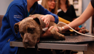
In veterinary medicine, understanding the complexities of an animal’s urinary system is vital for both diagnosis and treatment. One of the key...