Welcome to another informative radiograph review from SVS Imaging! In this post, we will delve into two important pathologies commonly observed in veterinary radiographs: right-sided heart enlargement and a diffuse bronchial interstitial pattern. Understanding these patterns is essential for diagnosing underlying conditions and providing the best possible care for our animal patients.
Radiographic Presentations
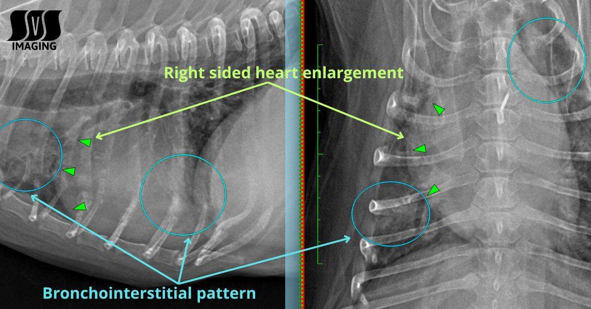
Right-Sided Heart Enlargement
When examining a radiograph, one of the first things to check is the size and shape of the heart. Right-sided heart enlargement is a common finding and can indicate a variety of conditions that affect the heart and lungs.
On the lateral view, the green triangles mark the borders of the right side of the heart, showing the enlargement. This enlargement can be a sign of several pathologies, such as valvular disease or pulmonary hypertension. In some cases, the enlargement may be subtle, but when it appears, it’s important to consider it as a part of the diagnostic puzzle.
Moving on to the ventrodorsal (VD) view, we observe a distinct bulging on the right side of the heart. This bulging further supports the possibility of right-sided heart enlargement and can provide valuable insight into the nature and extent of the issue.
Diffuse Bronchial Interstitial Pattern
Next, let's discuss the diffuse bronchial interstitial pattern. This pattern, which is indicated by the blue circles on both the lateral and VD views of the radiograph, is often seen in cases of chronic bronchitis, pulmonary infiltrates, or pulmonary hypertension. It can present as a subtle, diffuse haze on the image, with changes in both the bronchial and interstitial regions of the lungs.
This pattern may be associated with pulmonary hypertension, a condition that often occurs secondary to right-sided heart enlargement. Chronic bronchitis can also manifest this pattern, contributing to airway inflammation and thickening, leading to poor airflow and compromised lung function.
Primary Differentials to Consider
When interpreting radiographs with these findings, it’s crucial to form a differential diagnosis to guide the next steps in treatment. For this case, the primary differentials to consider include:
- Valvular disease of the right side of the heart – This condition can lead to heart enlargement due to increased pressure in the heart chambers.
- Pulmonary hypertension with secondary chronic bronchitis – Pulmonary hypertension often leads to changes in the lungs, including bronchial and interstitial patterns.
- Pulmonary infiltrates secondary to non-cardiogenic pulmonary hypertension – Infiltrates can be a result of pressure changes in the lungs, independent of the heart's function.
Why Early Diagnosis Matters
Detecting conditions such as right-sided heart enlargement and diffuse bronchial interstitial patterns early is essential for providing the best care for our veterinary patients. Understanding the underlying cause helps in deciding the most effective treatment options, whether it's managing heart disease, reducing inflammation in the lungs, or addressing any systemic effects.
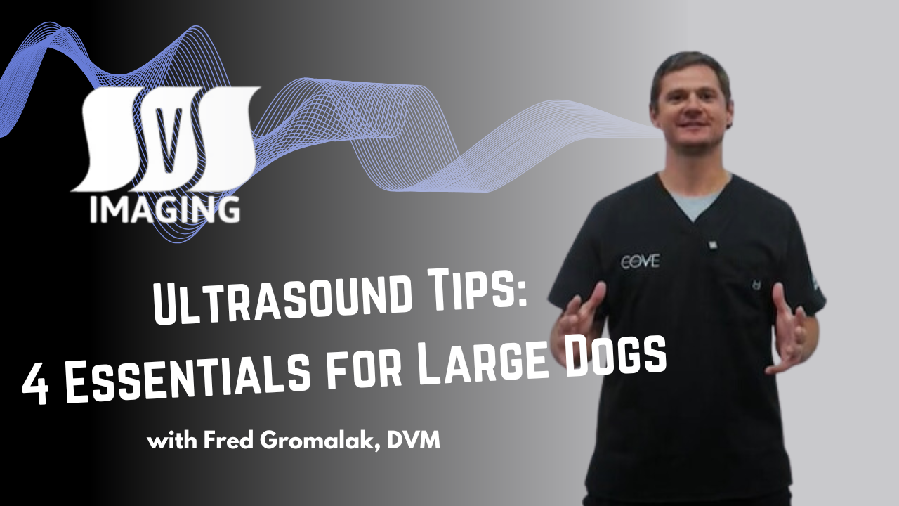
Watch the Video
Ready to sharpen your diagnostic skills? Watch the second episode of Reading with Dr. Fred: Understanding Right-Sided Heart Enlargement with Bronchial Interstitial Pattern
Need Help Interpreting Your Images?
If you’re looking to enhance your team’s radiographic interpretations or need expert assistance in reading your radiographs, SVS Imaging is here to help. Our teleradiology services provide fast, reliable, and detailed interpretations by board-certified veterinary radiologists, ensuring you get the most accurate diagnosis every time. Whether you're dealing with complex cases or need an extra set of eyes, we’re ready to support your practice.
- Click here to get a FREE teleradiology report from SVS Imaging.
*Offer valid for first-time customers only. Some conditions apply.
.jpg) Fred Gromalak, DVM
Fred Gromalak, DVM