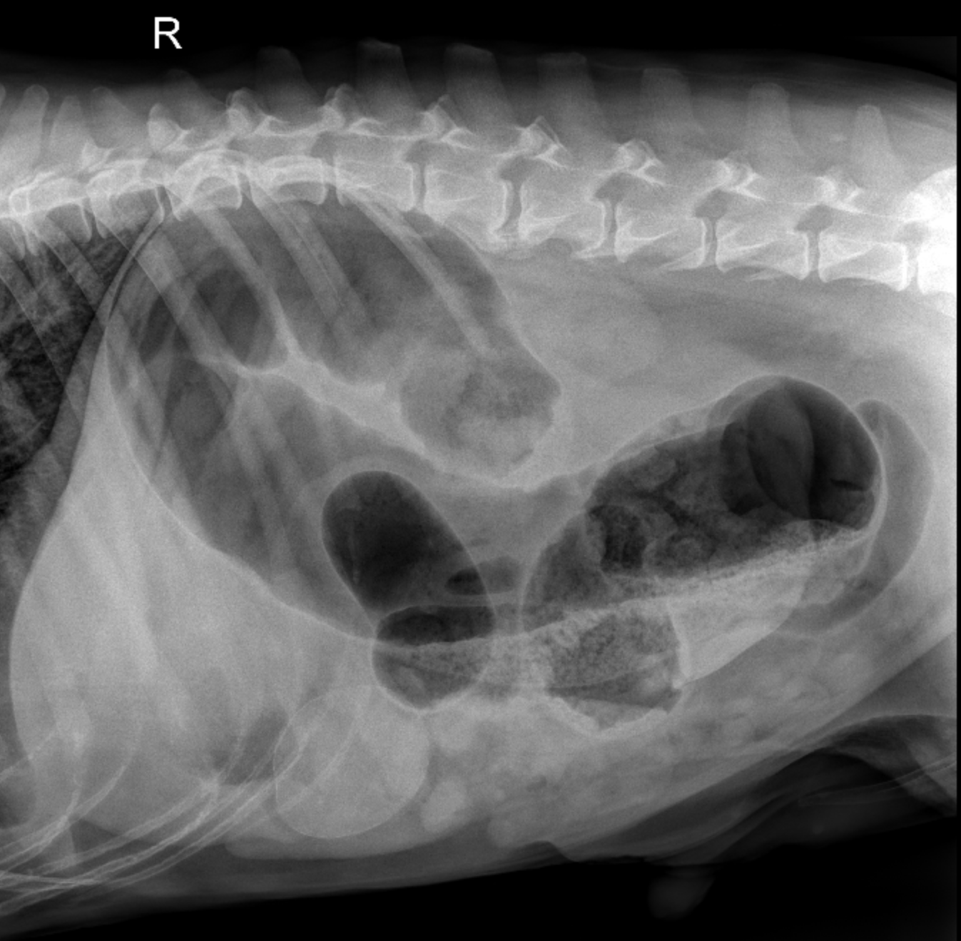
Radiographic Features of Colonic Torsion in Dogs
In this blog post, we’ll walk through the hallmark radiographic features of colonic torsion in dogs and why early diagnosis is essential for...
1 min read
.jpg) Fred Gromalak, DVM
:
Jan 23, 2024 12:34:20 PM
Fred Gromalak, DVM
:
Jan 23, 2024 12:34:20 PM

In today's video, we meet a 13-year-old male neutered border collie mix who has been dealing with chronic coughing and a noticeable decrease in appetite.
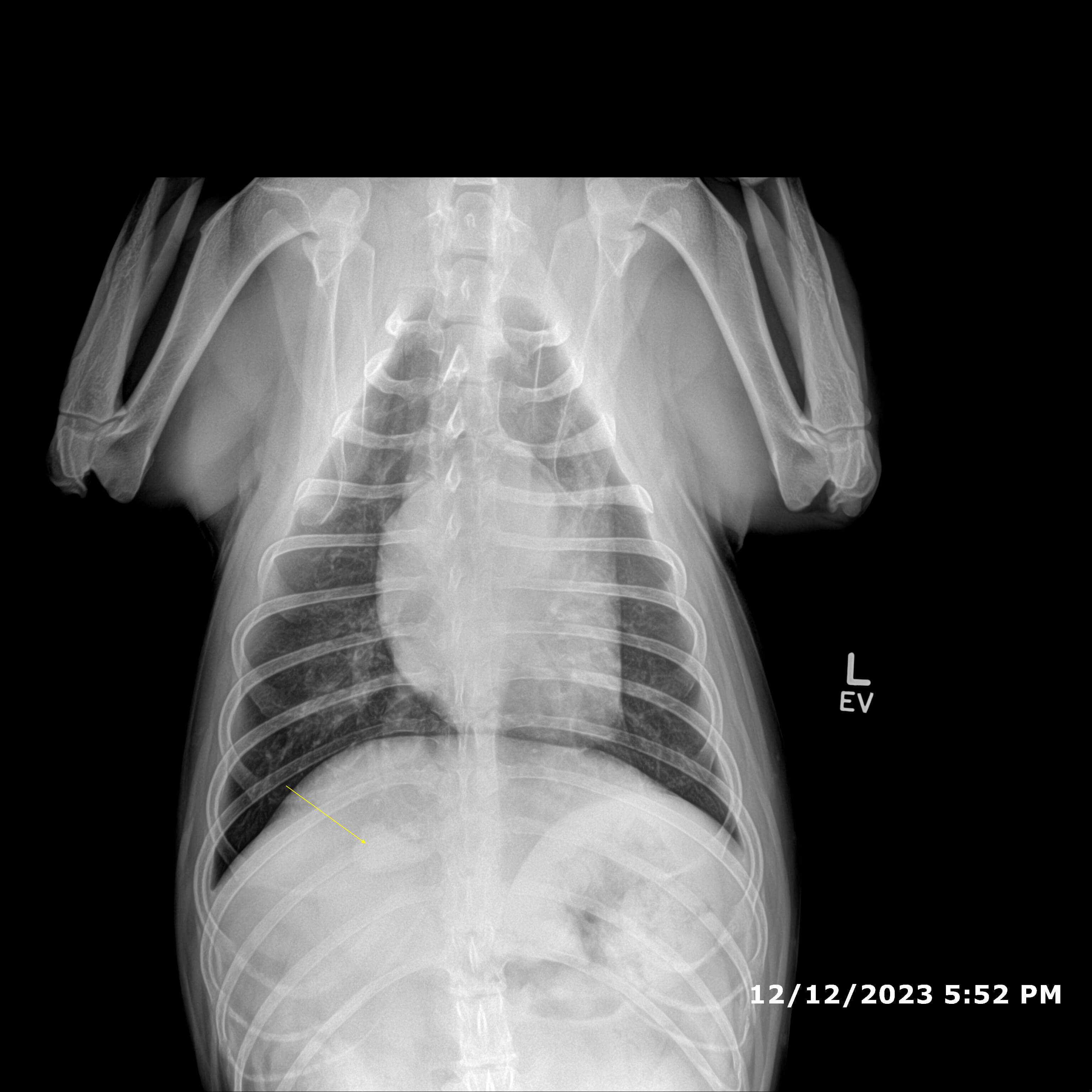
During our radiology review, our radiologist identified a large, soft tissue opacity mass located in the right hemithorax, specifically in the region of the right middle lung lobe. This mass exhibited mostly well-defined margins and had a significant impact on the dog's health. It caused compression of the cardiac silhouette, leading to a slight shift to the left side.
Our experienced radiologist considered several potential diagnoses for this concerning mass, with neoplasia, such as carcinoma, being one of the primary suspects. While other possibilities like cysts, granulomas, or abscesses were considered, they were deemed less likely.
To further investigate this health challenge, we discussed the next steps in the diagnostic process, which include a Fine Needle Aspiration (FNA) of the mass or more advanced imaging with a CT thorax. In some cases, a thoracotomy may be necessary to provide a definitive diagnosis and initiate appropriate treatment.
Get a FREE teleradiology report from SVS Imaging.
*Offer valid for first-time customers only. Some conditions apply.

In this blog post, we’ll walk through the hallmark radiographic features of colonic torsion in dogs and why early diagnosis is essential for...

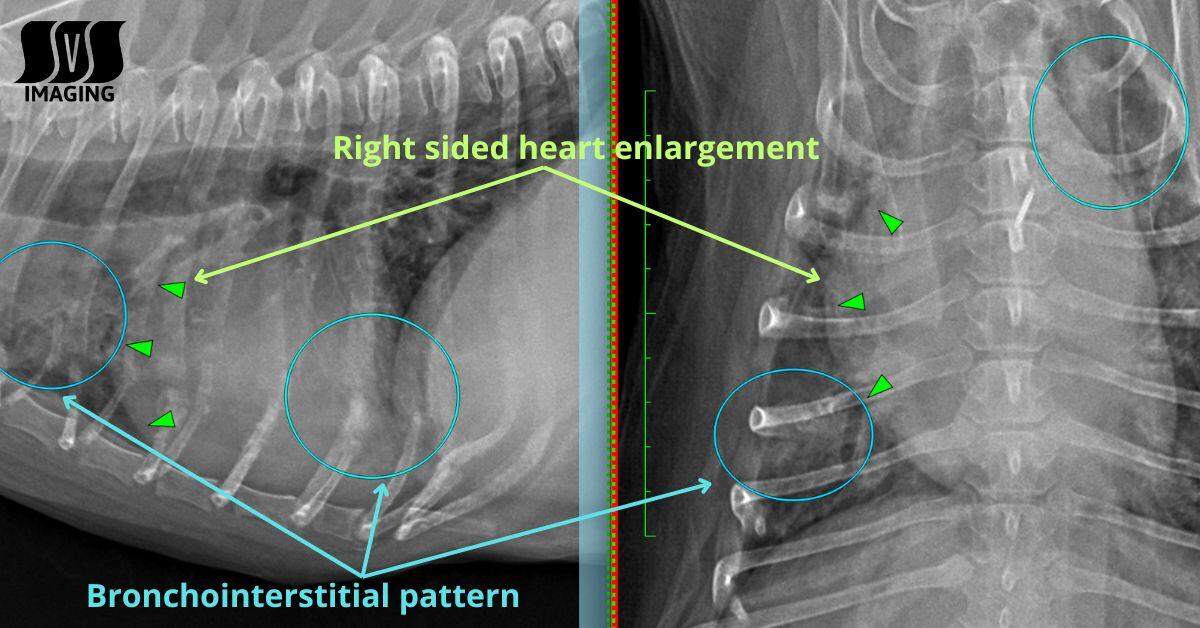
Welcome to another informative radiograph review from SVS Imaging! In this post, we will delve into two important pathologies commonly observed in...

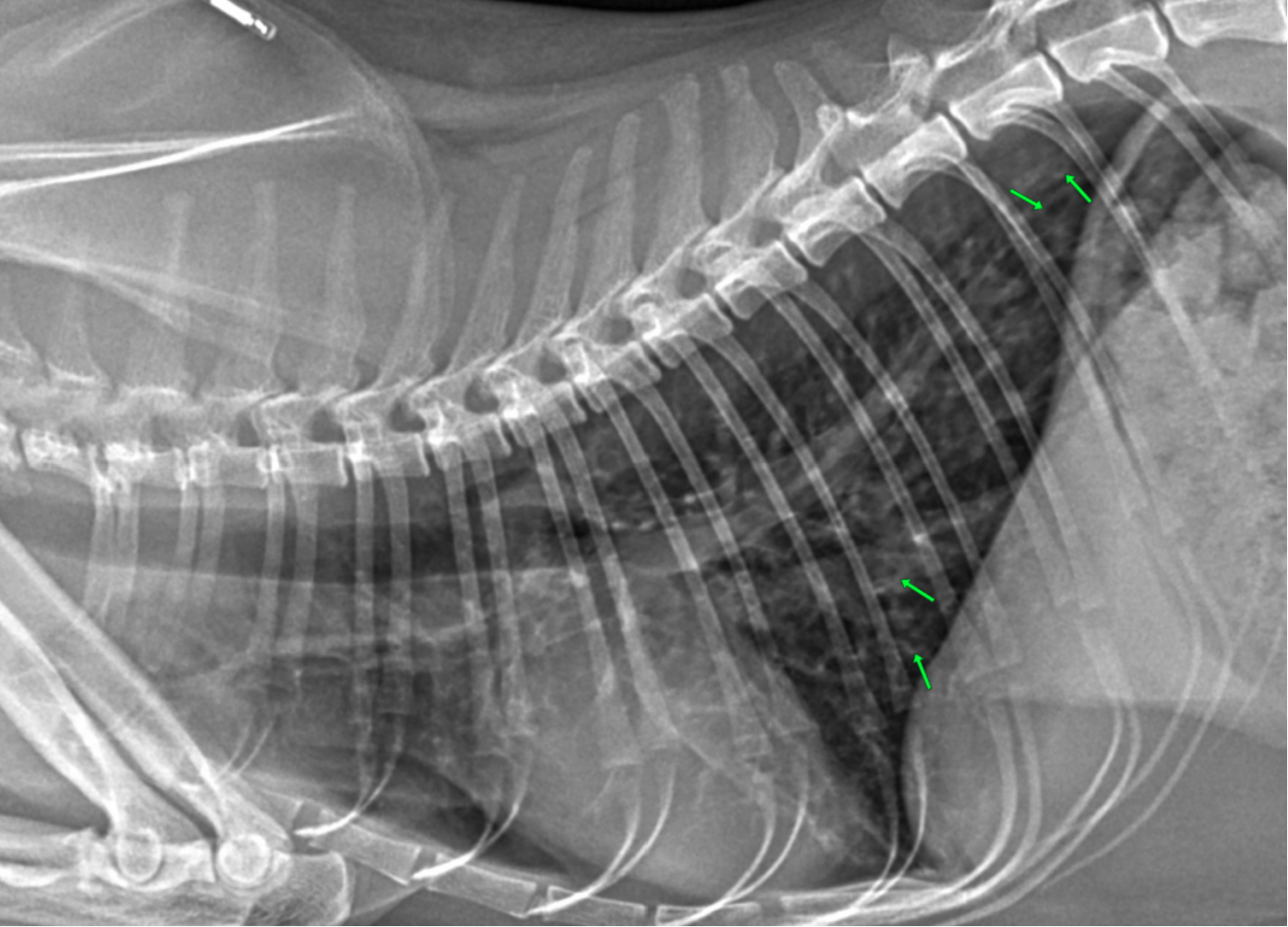
A Guide for Beginners