A Guide for Beginners
Radiographic imaging is a valuable tool in veterinary medicine, allowing us to visualize internal structures and diagnose various conditions in animals. However, it is important to understand and avoid common errors that can occur during radiographic examinations.
This guide will provide you with essential tips and techniques to help you minimize errors and obtain accurate radiographic images.
Positioning and Technique
Collimation and Centering
- Collimation restricts the X-ray beam to the desired area, reducing scatter radiation and improving image quality.
- Always center the X-ray beam on the area of interest to ensure optimal image clarity.
- Avoid excessive collimation that may crop out relevant structures or pathologies.
Technique Factors
- Select appropriate technique factors (kVp, mAs) based on the patient's size and the body part being imaged.
- Utilize techniques recommended by the equipment manufacturer or follow established guidelines.
- Adjust exposure factors to balance image quality and radiation dose.
Image Evaluation
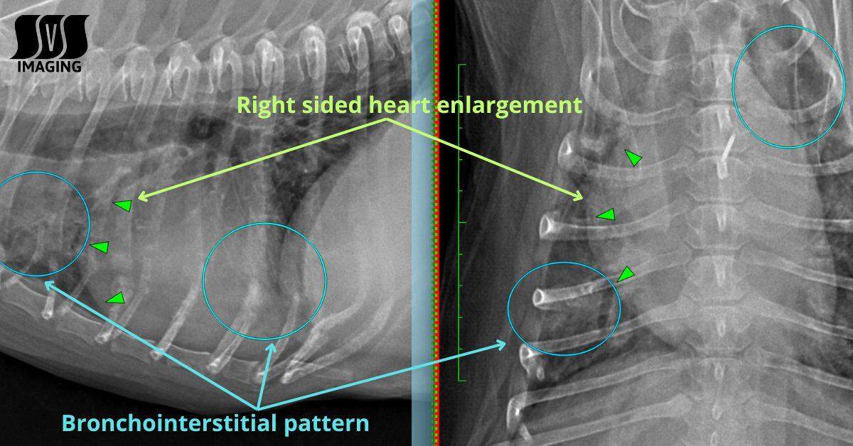
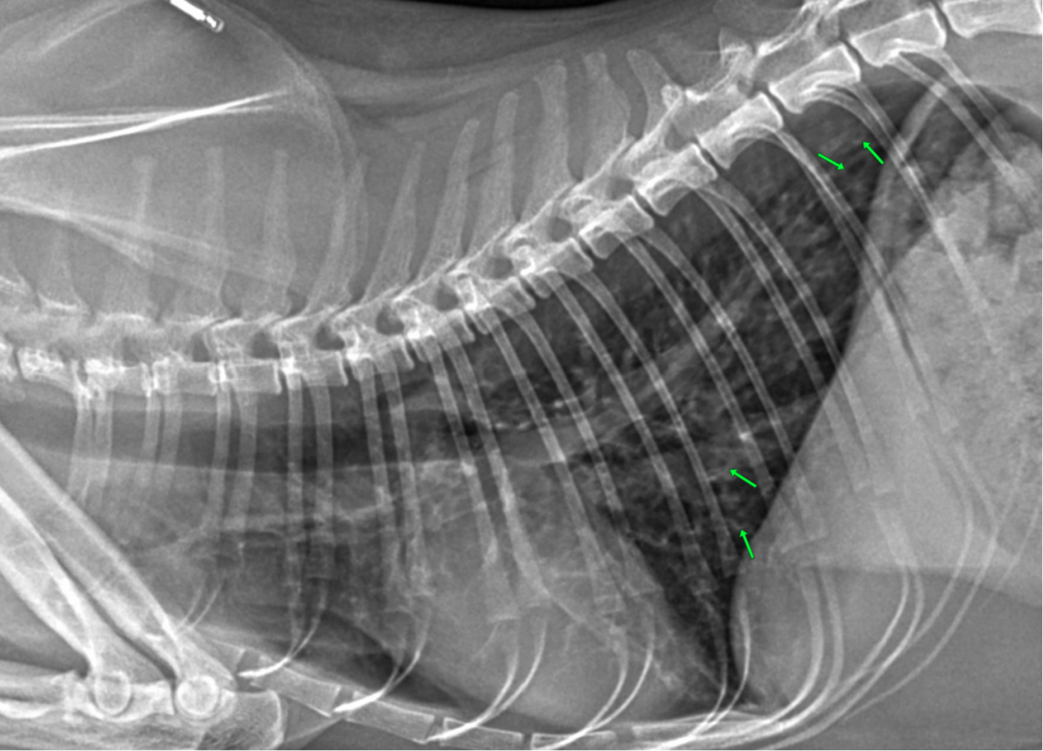
Artifacts and Error Identification
- Understand common artifacts that may affect image interpretation, such as motion blur, grid cutoff, or foreign objects.
- Identify and troubleshoot errors promptly to avoid misinterpretation or unnecessary retakes.
- Consult with experienced professionals or radiologists for assistance when needed.
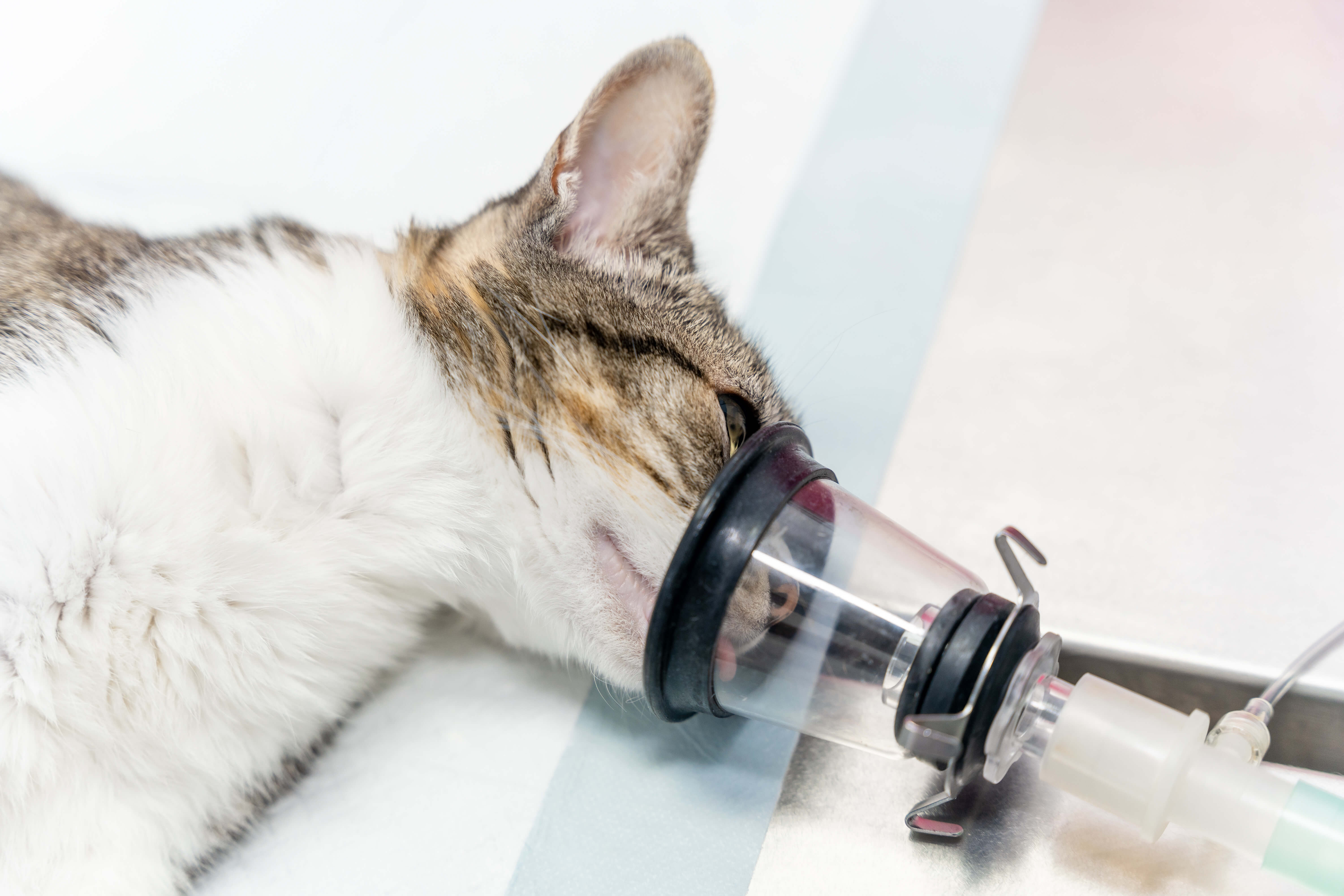
Patient Preparation and Safety
Radiation Safety
- Implement proper radiation safety measures for both patients and personnel.
- Use appropriate radiation protection equipment, such as lead aprons, gloves, and thyroid shields.
- Minimize radiation exposure by adhering to the ALARA (As Low As Reasonably Achievable) principle.
Correct Patient Identification
- Verify patient identification before performing radiographic examinations to avoid mix-ups or incorrect interpretations.
- Utilize identification tags, microchips, or other methods for accurate patient labeling.
Continuous Learning and Collaboration
Continuing Education
- Stay updated with advancements in radiographic techniques, equipment, and image interpretation through continuous learning.
- Attend conferences, workshops, or webinars to enhance your skills and knowledge.
Avoiding common radiographic errors is essential for accurate diagnosis and effective patient care in veterinary medicine. By following proper positioning and technique, evaluating image quality, prioritizing patient safety, and embracing continuous learning, you can minimize errors in veterinary radiography, ensuring accurate diagnoses and improved care. Prioritizing patient safety, continuous learning, and seeking collaboration elevate standards, benefiting the animals we serve. Dedication to detail and ongoing education are pivotal for success in this field, advancing veterinary medicine and enhancing patient outcomes.
Want to learn more?
Uncover crucial chest X-ray pathologies and diseases often missed. Download and print this checklist for your team to use as a reference.
Grab your free downloadable checklist now!
.jpg) Fred Gromalak, DVM
Fred Gromalak, DVM


.png)