In this blog post, we’ll walk through the hallmark radiographic features of colonic torsion in dogs and why early diagnosis is essential for effective intervention.
Understanding Colonic Torsion in Dogs: Radiographic Features and Diagnosis
When a colonic torsion appears on a radiograph, recognizing the key diagnostic features can make a critical difference for veterinary professionals. In the first episode of our new series, Reading with Dr. Fred, we dive into this uncommon but life-threatening condition to help veterinary teams identify it quickly and act decisively.
What is Colonic Torsion?
Colonic torsion, also known as volvulus, occurs when the colon twists around itself, obstructing the flow of gas and fecal matter. This twisting not only blocks the digestive tract but also cuts off blood supply, leading to tissue necrosis and severe systemic complications if untreated.
Key Radiographic Features of Colonic Torsion in Dogs
-
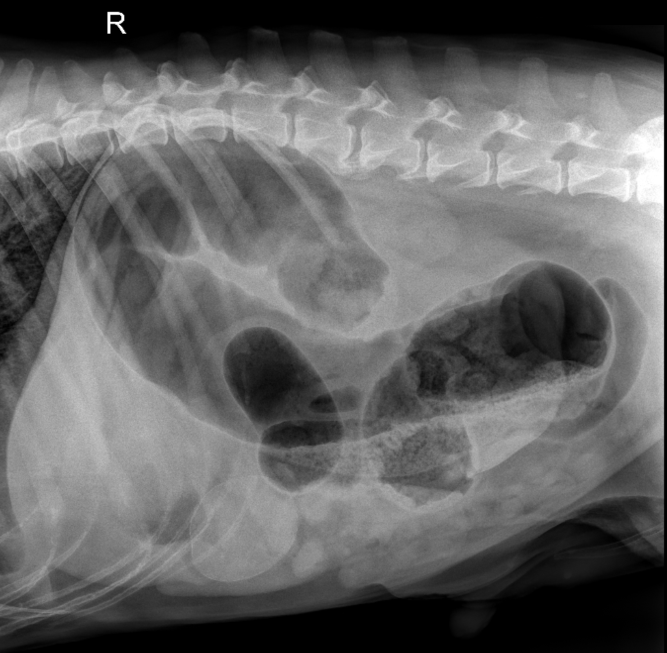
Large Gas-Distended Colon
One of the most significant indicators of colonic torsion is the presence of a gas-distended colon that forms a distinctive corkscrew or spiral shape. This is typically located in the mid-abdominal region, making it a hallmark sign on radiographs.
-
Tapering or Absence of Gas in the Descending Colon
Another critical feature to look for is a dramatic tapering or absence of gas continuing into the descending colon and rectum. This abrupt gas cutoff strongly suggests the presence of torsion, as the normal flow through the colon is disrupted.
By focusing on these two radiographic features, veterinary teams can confidently identify colonic torsion and begin the necessary interventions without delay.
Why Early Diagnosis Matters
Colonic torsion in dogs progresses rapidly, with severe implications for the patient. Delayed diagnosis and treatment can result in:
- Intestinal necrosis
- Septic shock
- Multi-organ failure
Quick identification of these radiographic signs allows for early surgical intervention, which is often the only treatment option to save the animal’s life.
Watch the Video
Ready to sharpen your diagnostic skills? Watch the first episode of Reading with Dr. Fred: Radiographic Features of Colonic Torsion in Dogs.
Need Help Interpreting Your Images?
- Click here to get a FREE teleradiology report from SVS Imaging.
*Offer valid for first-time customers only. Some conditions apply.
.jpg) Fred Gromalak, DVM
Fred Gromalak, DVM